


Image Computation for Microscopy
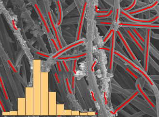
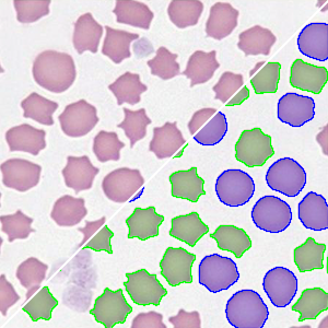
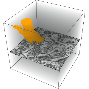
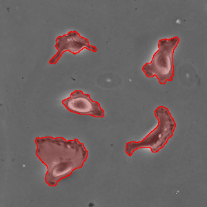
Version 12 adds new image processing functionality designed for specific tasks in microscopy, including biological sciences, material sciences, quality control and forensics. The capabilities range from brightness equalization to focus stacking and 3D reconstruction. In addition, with built-in machine learning and neural networks, the Wolfram Language is now the only high-level environment that provides advanced, state-of-the-art segmentation, feature extraction, object detection, classification and many more.
- Import of data and metadata from microscope-specific formats. »
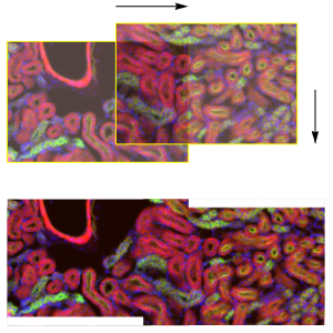
- Interactive functions to look into details of large images. »
- Comprehensive set of denoising and deconvolution algorithms. »
- Efficient brightness equalization to correct for uneven lighting. »
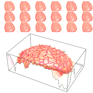
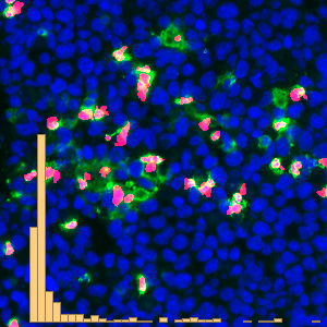
- Quantitative color analysis and visual enhancements. »