
")


Analyseskript zur früheren Erkennung von Herzunterschieden bei Neugeboreren und Erwachsenen
Ph. D. Craig Clarkson, Ph. D. William Crumb; Tulane University School of Medicine
Data analysis scripts played a critical role in a recent study investigating the physiological differences of heart tissue obtained from newborn and adult patients. The authors found significant differences in the behavior of electrical currents which help regulate the beating of the heart. The information provided by this study, and future studies, will help physicians decide which drugs, and drug dosages would provide the most beneficial effect for treatment of cardiac arrhythmias in newborns. The unique features of Origin software played a critical role in this study by greatly speeding up the rate of data analysis.
Drugs that produce their effects by binding to ion channels in the cell membranes of heart tissue are a primary treatment for potentially life-threatening cardiac arrhythmias. These drugs are commonly prescribed for adults who have heart disease brought on by such factors as age, smoking and diet. In addition, they are also commonly prescribed for treating arrhythmias in infants who have arrhythmias related to congenital heart defects, or by trauma to the heart created by open heart surgery designed to correct such life-threatening defects. Considerable knowledge is available on the action of these drugs on the hearts of adults. However questions have been raised as to whether there are significant physiological differences in the hearts of newborns which result in an altered sensitivity of newborns to the actions of these drugs. This question was recently addressed by scientists at the Tulane University School of Medince, who performed a study designed to compare the physiological properties of two electrical currents in the human heart, the transient outward current (Ito), and the inward rectifier (IK1). These currents, which play an important role in regulating the beating of the heart, are both produced by the flow of charged potassium ions through proteins in the cell membrane which function as ionspecific channels or pores. Based upon clues provided by previous studies in rats, dogs and rabbits, these investigators were looking for significant age-related differences in the properties of these channels in the human heart.
The investigators carried out their study on specimens of human heart tissue obtained from patients undergoing open heart surgery. The tissue was obtained in accordance with guidelines established by the Tulane University School of Medicine. During open heart surgery patients are temporarily connected to a blood gas machine (pulmonary bypass) that reoxygenates venous blood and then pumps it into the arterial circulation. This allows surgeons to repair defects or damage to the heart while maintaining a constant flow of blood to other vital organs, such as the brain and kidneys. Typically this machine is attached to the patient by tubes which are placed into small holes cut into the right atrium and aorta. To cut a hole in the atrium, surgeons typically lift up and clip off a small piece of the atrium about the size of a pencil eraser. This tissue, which is normally discarded, can be collected and taken back to the laboratory for study. In this recent study Tulane scientists used such samples of human heart tissue to isolate individual heart cells which were then placed into a tissue bath for analysis.
Heart cells obtained from 20 newborn patients (ages 1 day - 2.5 years) and 8 adult patients (ages 11-68 years) were included in the study. The electrical currents in these single heart cells were measured under a variety of experimental conditions utilizing a highly sensitive recording amplifier (Axon Instruments, Inc., Foster City CA). The electrical signals detected by this amplifier were digitized using an analogto-digital converter and stored on a personal computer in a binary file format. In most experiments, hundreds to thousands of data traces were stored for later analysis. To analyze the large amount of data obtained during each experiment, each data file had to be imported into one (Axon Instruments) program which could detect peak values. The peak signals for the hundred or more data traces recorded during an experiment would then be imported into one or more additional programs for additional analysis, curve fitting, and statistical analysis.
Typically, due to incompatibilities in import/export features between different programs, peak values had to be written down on paper and then manually typed into a spreadsheet table in each additional program. This timeconsuming, and often monotonous task of peak detection, data transcription, curve fitting, and statistical analysis for hundreds of data values typically required a full day for analysis of data collected during a one hour experiment.
Tiring of this task, Tulane scientists searched for a more robust data analysis package that would allow them to do everything needed, such as peak detection, curve fitting, statistics, and production of hard copies of both data values and electrical recordings in one software environment. To accomplish this goal, they selected Origin data analysis software from OriginLab, Corp., Northampton, Massachusetts. Two reasons for selecting this software were its ability to import data stored in the Axon binary file format, and the powerful Labtalk scripting language that can be used to produce a highly automated, customized and user-friendly data analysis program.
The first task in creating this data analysis program was to read and import the binary data stored from the Axon Instruments amplifier. This was accomplished using an Origin add-on module designed for importing data stored in the Axon binary file format. This "pCLAMP" module allowed for easy access and plotting of the experimental data, as well as access to a variety of data header information stored at the beginning of each Axon binary data file.
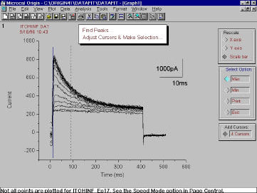
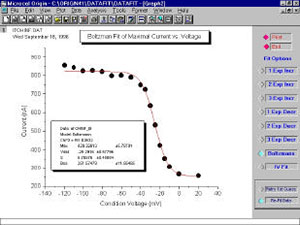
While Origin comes supplied with a built-in peak detection routine, Tulane scientists preferred to develop a customized script-driven graphical interface that would enable a visual confirmation of the peak detection process. To initialize the peak detection routine, the user first imported and plotted a series of experimental data traces in a graph displayed on the PC monitor. Then using a mouse, the user placed two vertical cursors (vertical colored lines) on each side of the range of peaks displayed on the graph, and clicked a user-designed button to activate a script which would search for peak values in each data trace at positions between the two cursors. When all peak values were determined, the script stored the peaks as "Y" values in a table and associated each of these values with an appropriate "X" value determined from analysis of the header information stored in the binary file. When all X and Y values were determined, the script would generate a new window displaying a graph of the X vs. Y values, and a series of buttons off to one side. Selection of one of these buttons by a mouse click allowed the user to select from a variety of different types of user-defined fitting functions, such as an increasing or decreasing exponential, linear function, sum of exponential functions, or a Boltzmann function. When selected, the button would activate a script which would calculate a least-squares fit of the selected equation to the experimental data, and display both the best-fit, and the best-fit values. An additional button, when selected, would send a hard copy of the data fit, least-squares fit parameters, and a print out of the X and Y values, to a laser printer.
Since each step in the data analysis was fully integrated, the entire process of importing data from the Axon binary file to print out of the final stage of data analysis required less than 2 minutes for a set of 15 to 50 data traces, a greater than 5 to 10 fold reduction in the amount of time it would take to analyze the same data using a combination of different programs. As a result, Tulane scientists were able to collect, analyze, and submit for publication a large amount of experimental data in a fraction of the time it would have taken using previous methods.
The end result of their recent study on human heart tissue was that there are significant age-related changes in the properties of the transient outward current (Ito), including: 1) the absence of the Ito in 33% of all cells from newborn hearts, in contrast to an incidence of 100% in adult cells, 2) a 2-fold lower amplitude of Ito in heart tissue from newborns compared to adults, and 3) a significant age-related change in the kinetics with which the channels switch from closed to open states in-between heart beats. These results indicate that there are important age-related changes in ion channels that occur in the human heart. Since ion channels are the site of action for many of the drugs used to treat cardiac arrhythmias, this study suggests that there may be significant differences in how the newborn heart responds to drugs that act by blocking these ion channels.
Tulane scientists are currently working on expanding the Origin-based data analysis program to include a final phase of statistical analysis of the data. This script-based analysis will determine mean values for fit parameters obtained by analysis of multiple cells from patients in different age groups, and perform group comparisons using standard statistical tests such as Student's t test, and analysis of variance (ANOVA). Once these script routines are completed, all phases of data analysis, except for data collection, will be completed in one seemless and integrated software environment.